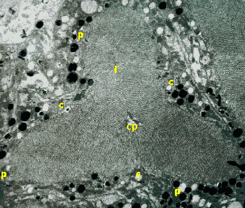
Here we are looking at cells in the eye of Opilio parietinus. This harvestman belongs to a diferent family from our other favourite, Mitopus morio, but the general structure of its eye is similar.
In the phalangid eye, the shape of the rhabdoms, as seen in cross-section,
varies between species and across the retina of individuals. In this image,
we can see a cross-section of a retinula showing the rhabdom (r) with central
process (cp).
The distribution of the central (c) and peripheral (p) cells is evident.
The arrangement of these in the structure of
the retinula is the same as in Mitopus morio.
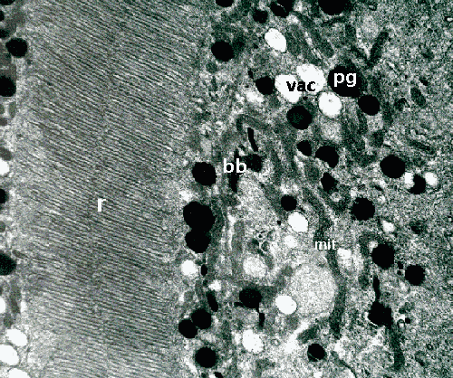
This plate illustrates the structure of the rhabdom (r), in addition to some of the organelles in the retinula cell cytoplasm. These include pigment granules (pg), vacuoles (vac), bizarre bodies (bb) and many peri-rhabdomeric vesicles (prv). The mitochondria are very long and often tortuous.
The overall appearance of the cytoplasmic organelles is denser than our observations on Mitopus morio, and it is interesting to speculate whether this respresents interspecific variation or different physiological states.
If you look in more detail, the Golgi complex is one of the very interesting organelles in these cells.
As shown in the mystery picture for August,
at the edges of the rhabdom of O. parietinus, there are lamellar
membranes underlying the microvilli. These lamellae are accompanied by
large numbers of peri-rhabdomeric vesicles. These can be clearly seen down
the right-hand side of the image, which is close to the edge of an adjacent
rhabdom.
For further details, see:
Curtis, D.J. (1970) Comparative aspects of the fine structure of the
eyes of Phalangida (Arachnida) and certain correlations with habitat. J.
Zool., Lond. 160: 231-265.
|
Back to Opilio Notes | or | Back to Arachnologia |
 |
Back to Home page |