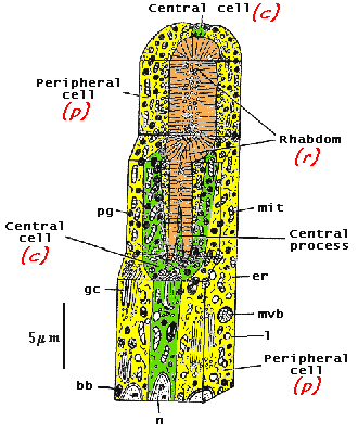
Diagram of a retinula. |
Separate out the cells again. |
1. Use this button to move the rhabdom (r) into place on the central cell (c). |
2. Then you can close in the peripheral cells (p) with this button. |
|
 |
 |
|||
Diagram of a retinula. |
Separate out the cells again. |
1. Use this button to move the rhabdom (r) into place on the central cell (c). |
2. Then you can close in the peripheral cells (p) with this button. |
|
|
|
|
|||
| Back to:- | ||
| The mystery picture | Opiliones Notes |
Arachnologia |
 Ariadne
Home Page Ariadne
Home Page |
||