

| Back to Opiliones Notes |
|
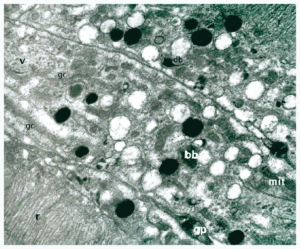
Here we are looking again at cells in the eye of Opilio parietinus.
The cytoplasmic contents of M. morio appear quite sparse, with the organelles showing up very clearly, epecially the long, almost filamentous mitochondria and the very dark pigment granules. In contrast, as you can see here, in the retinula cells of O. parietinus the cytoplasm appears much denser. In this image we can see part of two rhabdoms (r) and three adjacent retinula cells. In these, as in M. morio, mitochondria (mit) and pigment granules are evident, but there are also lots of fairly large vacuoles with clear contents. More strikingly, the rough endoplasmic reticulum is more plentiful than in M. morio and there are large numbers of vesicles (v) of moderate electon-density and other organelles which are very opaque to electrons - dense bodies (db) and bizarre bodies (bb). In between the retinula cells, even the glial processes (gp) have dense contents.
Another feature of particular interest in these cells are the large Golgi complexes (gc). Note: the "closer look" is not just an enlargement of part of the first image. It is from a micrograph taken originally in the electron microscope at higher magnification.
The overall appearance of these organelles (and of the whole cell contents) is suggestive of a high level of metabolic activity. Remember that a cell is not just a static structure - it is a whole set of interacting functional processes interdependent with the cell's structures. Materials don't just have a fixed location within the cell - rather there is a movement around and through the cell as structures change position or as transport processes take substances from their site of synthesis to their site of function. As shown in the mystery picture for August, at the edges of the rhabdom of O. parietinus, there are lamellar membranes underlying the microvilli. These lamellae are accompanied by large numbers of peri-rhabdomeric vesicles which can be seen at the edges of the rhabdoms at top-right and lower-left of our image here. The materials for these vesicles are synthesised elsewhere and then transported to their final site of functioning.
The term "endoplasmic reticulum" translates roughly as "network inside
the cytoplasm" and well describes the rambling of membranes throughout
the interior of the cell. It is easy to imagine materials being transported
along (or in, or by) these membranes; this is particularly the function
of the smooth endoplasmic reticulum, while the rough endoplasmic reticulum
(studded with ribosomes) is the location for synthesis of proteins.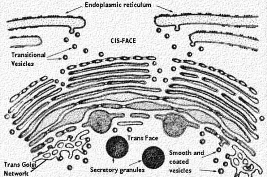
The Golgi complex is a specialised part of the endoplasmic reticulum
engaged in the processng and transport of materials.
As you can see in this graphic, vesicles come off from the endoplasmic
reticulum and enter the Golgi complex at one side - called its cis-face.
These transitional vesicles are taken up by the membranous lamellae of
the Golgi complex and emerge from the other side (the trans-face) transformed
into products with particular functional roles: secretory granules, coated
vesicles, etc.
Want to know more? .... Just click on the image to go to the University
of Arkansas and visit Gwen
Childs informative site.
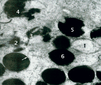
As you can see, the pigment granules of the retinula cell cytoplasm contain varying amounts of electron dense material, ranging from almost completely empty (1) to completely filled (6), and it is possible to trace a series of increasing contents (1 to 6) possibly representing a developmental process.
The pigment granules are bound by a membrane which may sometimes be
seen to turn away from the organelle (at the arrow) and join the endoplasmic
reticulum. Also check out the mystery picture
for February.
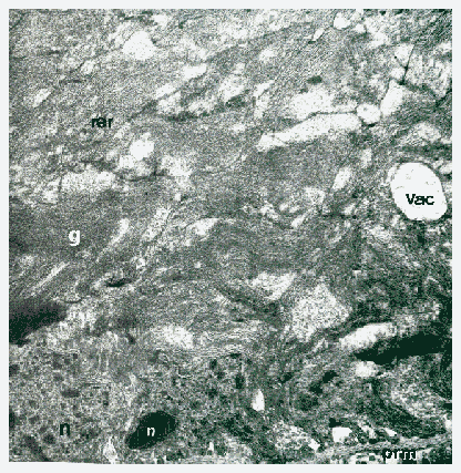
Close to the pre-retinal membrane (prm) we can see the nuclei
(n), in which there are many dense areas. There are some relatively
clear ares in the cytoplasm of the lentigen cells and fairly large emplty-looking
vacuoles, but the bulk of the cytoplasm is packed with dense granular material
(g) and masses of reough endoplasmic reticulum (rer).
|
Back to Opilio Notes | or | Back to Arachnologia |
 |
Back to Home page |