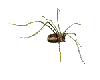

 If
you look sideways at a harvestman - no, that's not quite right! - if you
take a lateral view of a harvestman, as shown here, you can see that there
is a single pair of eyes mounted dorsally on its back, forming a distinctive
structure termed an ocularium.
If
you look sideways at a harvestman - no, that's not quite right! - if you
take a lateral view of a harvestman, as shown here, you can see that there
is a single pair of eyes mounted dorsally on its back, forming a distinctive
structure termed an ocularium.
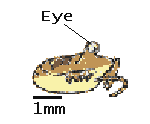
Rather a strange place to have your eyes - it probably means that you
can't look where you're going, but that doesn't matter to harvestmen as
they get their main sensory input through their long legs (especially the
second pair).
However, these eyes are well developed, each with a clearly
evident large lens, beneath which is a retina made up of sensory units,
called retinulae, packed together.
It is interesting to note that the retinula cells conatin many mitochondria. The mitochondria are the structures which provide the cell with its energy requirements and the presence of lots of these indicates that these cells are very active.
In addition to these retinula cells, which are essentially highly modified sensory nerve cells, in the retina there are glial cells which appear to have a supportive function.
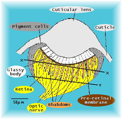
At the side of the eye are pigment cells,
preventing the entry of stray light and restricting the incoming light
to rays passing through the lens.
Close examination of the retinulae show that each is made up of four cells. There are three peripheral cells which extend unbroken up the side of the rhabdom. The fourth, or central, cell divides into three parts which extend up between the peripheral cells, as well as a central process which projects into the base of the rhabdom.
The pattern of these peripheral and central cells is shown in these
diagrams of cross-sections.
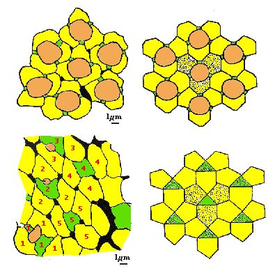
The drawings with the size scales are taken from electron micrographs;
the diagrams to their right show the geometrical basis of the arrangement
of these cells. Stippling emphasises the structure of the retinulae.
The upper drawings show a transverse section
through the rhabdoms, with the peripheral cells cooured yellow and the
central cells green. You can see how the mall processes of the central
cell are interposed between the peripheral cells. The rhabdoms are shaded
a sort of orangey-pink (on my screen anyway) as an allusion to the visual
pigment in their membranes; this pigment is retinal, which is chemically
related to vitamin A and to carotene (the red colour of carrots (Bugs Bunny
does
have good vision!).
This corresponds to a section through the retina (marked X-X) in the
diagram of the eye, as illustrated in the mystery
picture for December.
The lower drawings are lower in the retina, below the level of the rhabdoms
(Y-Y in the eye diagram), but with a few rhabdom bases showing in retinulae
1, 2 and 3. The cells in five of the retinula are numbered to indicate
the cellular arrangement.
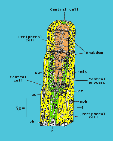
These cells are also indicated in the sketch of the three-dimensional
structure of a single retinula.
You can see how the central cell is positioned beneath the rhabdom;
it then has a short conical central process in the base of the rhabdom
and the three proceses in between the peripheral cells. Two of the latter
are shown on either side of the rhabdom; the third lies beside the rhabdom
in the plane in front of your screen. This may be seen
more clearly in animation.
Some of the organelles which can be seen in these cells are shown:-
pg: pigment granules - black discoid structures which
prevent lateral passage of light through the eye; they give a black colour
to the retina (NOT the yellow, green and pink as shown here!).
mit: mitochondria - elongated, ovoid organelles
involved in the energy metabolism of the cell.
er: endoplasmic reticulum - two kinds, (a) rough,
sometimes with a lamellate appearance and always studded with bead-like
ribosomes (the sites of protein synthesis, and (b) smooth er, often tube-like
in appearance and lacking ribosomes but involved in transport of materials
around the cell.
gc: Golgi complex - comprises lamellar, tubular
and vesicular structures, involved in processing of cell secretory products,
etc.; so-called because they were originally seen by Golgi with his wonderful
staining method.
l: lysosome, relatively large vesicular structures in which
cell components are broken down by enzymic action (more about this later).
mvb: multi-vesicular body - a cluster of small vesicles packed
together; identified as a kind of lysosome.
bb: bizarre body - a darkly-pigmented structure
containing remnants of other cell organelles; another kind of lysosome.
n: nucleus - containing the chromosomes, holding
the genes which control the cell's protein synthesis (and hence its features
and properties); the genes carry information coded in DNA which controls
the RNA which determines the protein synthesis in the ribosomes of the
rough endoplasmic reticulum.
Most remarkably, my e.m. work confirmed observations made in the 19th
century! A significant paper is:
Purcell, F. (1894) Uber den Bau der Phalangidenaugen. Z. wiss. Zool.58:
1-53.
For further details, see:
Curtis, D.J. (1970) Comparative aspects of the fine structure of the
eyes of Phalangida (Arachnida) and certain correlations with habitat. J.
Zool., Lond. 160: 231-265.
|
Back to Opilio Notes | or | Back to Arachnologia |
 |
Back to Home page |