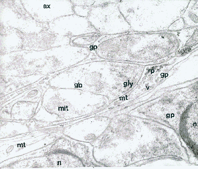
The retinula cells of the retina are, in fact sensory neurons and we can see their axons (ax) in this section located behind (or proximal to) the retina.
But also in evidence at this level in the retina are the cell bodes of the glia (gb). They contain a nucleus (n) and mitochondria (mit), but special features also occur which may be involved in a transport role of these cells, including vesicles (v), glycogen granules (gly) and particles thought to be ribosomal in nature (rp).
Like the glial processes (gp) the glial cell bodies contain microtubules
(mt). These microtubules are remarkable organelles, having a supportive
role equivalent to our skeleton, and also being capable of transporting
materials along their length.
We move further up into the retina for the next view of these cells.
This electron micrograph is of a section taken slightly further into
the retina, so that more of the retinula cells bodies show, including their
nucleus (n), as well as other organelles. They're not labelled,
but there are stretches of endoplasmic reticulum as well as detached ribosomes
and other particulate organelles. Amongst the most prominent features,
we can see one or two pigment granules appearing as the large, round, electron-dense
black structures but some of the less dense large round structure could
well be secretory in nature. There are also assorted vesicles and lysosomal
organelles as well, of course, as mitochondria.
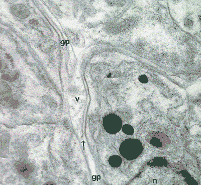
Also, in between the retinula cells, you can see the glial processes (gp), containing many vesicles (v). They also contain those marvellous microtubules, one of which is indicated by the arrow pointing along its axis.
Remarkably, when we look at the distal part of the retina, we can still see these glial processes which project along the full length of the retinulae.
The cell membranes appear very sharply here when they are cut cleanly
in transverse section (perpendicular to their surface, but they become
much more indistinct when cut obliquely or in a plane almost parallel to
their surface. This applies to all of the electron micrographs in this
series (and elsewhere).
For further details, see:
Curtis, D.J. (1970) Comparative aspects of the fine structure of the
eyes of Phalangida (Arachnida) and certain correlations with habitat. J.
Zool., Lond. 160: 231-265.
and
Curtis, D.J. (1969) The fine structure of photoreceptors in Mitopus
morio (Phalangida). J. Cell Sci., 4: 327-351.
|
Back to Opilio Notes | or | Back to Arachnologia |
 |
Back to Home page |