
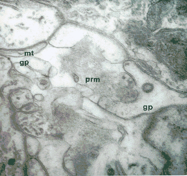
The section is cut just at a slight tangent to the pre-retinal membrane
(prm), which shows up as the rather amorphous material, but with
a finely filamentous texture.
Note that we can see the glial processes (gp) of the glial cells,
whose cell bodies are located in the proximal
part of the retina. Even at this distal level, these processes still
contain microtubules, one of which is labelled (mt). Remember that,
in looking at the proximal parts of the glial cells, we noted "...
microtubules are remarkable organelles, having a supportive role equivalent
to our skeleton, and also being capable of transporting materials along
their length." So, these glial cells can have a supportive role for
the full length of the retinula cells of the eye, both in a physical,
"architectural" sense and in a physiological sense supplying essential
needs.
The appearance of the other cells in this electron micrograph suggests
that two types are present:
Lentigen cells - at the lower left-hand corner of the image,
with some secretory material in evidence;
Retinula cells - at the upper right-hand corner, with more obvious
cell organelles including some endoplasmic reticulum and mitochondria.
I rather like this particular electron micrograph with its subtlety
of fine detail - so I've left it rather large, just reducing it to two-thirds
of its original size (in terms of pixels). It looks good, anyway, on my
screen at 1024 x 768 pixels.
For further details, see:
Curtis, D.J. (1970) Comparative aspects of the fine structure of the
eyes of Phalangida (Arachnida) and certain correlations with habitat. J.
Zool., Lond. 160: 231-265.
and
Curtis, D.J. (1969) The fine structure of photoreceptors in Mitopus
morio (Phalangida). J. Cell Sci., 4: 327-351.
|
Back to Opilio Notes | or | Back to Arachnologia |
 |
Back to Home page |