| Here is that electron micrograph again - it shows us features
of the cytoplamic organelles in the retinula cell of
Mitopus morio.
We can see vesicles (v) with those little dark spots. These marks are, in fact, an electron-dense stain produced by the action of the enzyme acetylcholine esterase (AChE). Also visible, between the dark masses of the pigment granules, are other cell organelles such as mitochondria and endoplasmic reticulum. Of greatest interest, though, is the relatively clear profile of a Golgi complex, with its stacked membranes and associated vesicles - some of which contain AChE (arrowed). |
|
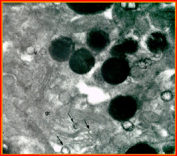 |
This, not surprisingly, is the connection with
September's structure - the Golgi complex
is of central importance in the cell's processing of materials.
We started off with an image of the rhabdom . If you look closely enough, you can see AChE
in the vesicles surrounding the rhabdom.
Of course, care is needed to verify that the staining is the result of the particular enzyme.Check out how we can use a specific inhibitor to do this. |