 In
the eye note:
In
the eye note:
With thanks to my colleagues, Robin Orchardson and Fred Toates.
Prof. Dave Curtis.
Our perception of the world depends upon a complex interaction between two things:
(1) raw sensory information (also termed `data') and
(2) the context, stored memories, expectations (also termed `concepts).
Thus, perception is said to be data driven and concept driven.
Two meanings of terms bottom-up and top-down:
(1) in the process of perception;
(2) in the process of scientific understanding. For example, an explanation
might be said to be sought by reduction and thereby given `bottom-up'.
You could start bottom-up in both senses of the meaning:
(a) by looking at the raw sensory data and how it is translated into
neural signals
and (b) by considering the insight into perception that can be gained
by looking at nervous system processes.
Consider the claim (Section 4.1) "...vision will be regarded as an indirect and active process which begins with a description of the image but which must also solve the formidable problem of working out what object produced the image'.
First stages of visual processing can be called descriptive and later stages interpretive.
Note that flat 2-dimensional image becomes interpreted as 3-dimensional
world.
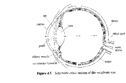
In
the eye note:
(1) cornea
(2) lens
(3) retina
(4) fovea
(5) optic nerve
(6) blind spot
(7) pupil
Note that retina is `inside out' and the reason why a blind spot exists.
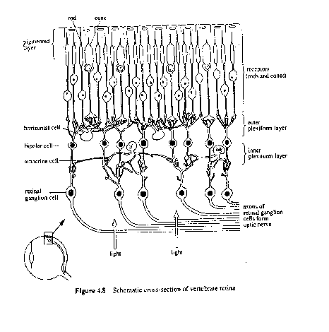
Graded potentials in cells at early stages - action potentials only
appear
at the ganglion level.
Note convergence of receptor cells on to retinal ganglion cells.
This convergence varies over retina. It is least at the fovea (shown
in first figure) = high acuity.
Retinal receptors contain rhodopsin.
This chemical interacts with light and in so doing the receptor cell
hyperpolarizes.
Note that this is the opposite of what normally happens in sensory
detection (e.g. depolarization of a nociceptor).
In the dark there is a certain background level of transmitter release.
Thus light absorption causes hyperpolarization causes reduction in
transmitter release.
Remember how to measure the receptive field? Check it out on the CD-rom, or in Book 3, Ch. 4.
There are two antagonistic regions of the receptive field: on-region
(light causes increase in firing rate) and off-regions (light causes decrease
in firing rate).
These regions form concentric regions:- on-centre and off-centre cells.
Study Note:
So what function do ganglion cells serve?
With the opposing areas in their receptive fields, they provide a way
of making a comparison - detection of edges, distinction between small
light object and large white object. It is interesting to compare this
with similar processes in the touch receptor system.
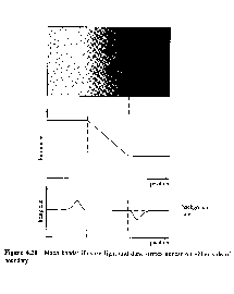
But ganglion cells also can play a role in the detection of luminance
discontinuities of the kind shown here (Figure 4.13 of Book 3).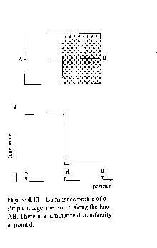
This is achieved as shown below, which should be compared with the schematic
shown in Figure 4.16 of Book 3. 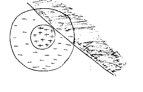
Here, the dark area covers part of the inhibitory surround of the receptive
field - so there is an increase in the cell's firing rate.
See Figure 4.21 on p.98. reproduced here.
However, note that the ganglion cell output explains part of your perception but not all of it.
You tend to see something like the profile shown in the figure below.
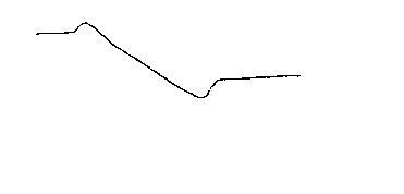
Hence it seems that the perceptual process extrapolates based upon information at luminance discontinuities.
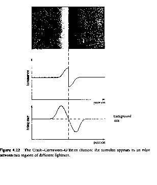
Evidence that this is so is provided by the perception of the figure
here (reproduced from Figure 4.22 of Book 4).
Processing of the visual information continues in the lateral geniculate
nucleus (LGN). 
The receptive field of an LGN cell is similar to that of a retinal ganglion
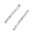
cell but cortical cells have slit-like, elongated receptive fields as shown
here.
These cells then project on to the visual cortex, where further processing takes place.
Note that receptive fields of cortical cells, as with the retinal ganglion
cells, are defined in terms of retinal surface and the positions of light
and dark parts of the image.
Various types of cortical cell:

1. simple cells -
one fixed position of optimal stimulus
2. complex cells -
receptive field such as to allow a variety of different
stimuli at a given orientation to trigger the cell.
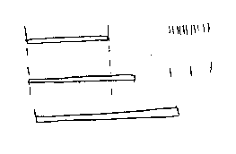
3. hypercomplex cells -
similar to cornplex but respond less if stimulus
protrudes beyond receptive field's positive area.
See Figure here (Figure 4.26 of Book 3)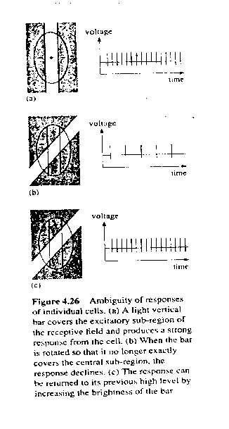
and compare with story of colour vision given later.
The individual cell portrayed in Fig. 4.26 cannot differentiate between
a dim stimlus aligned to its on-region and a bright stimulus at a different
orientation.
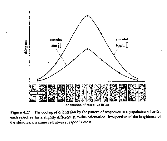
Note in Figure below (Figure 4.27 of Book 3) how the comparison is performed.
The figure shown here (after Figure 4.34 of Book 3) shows the responses 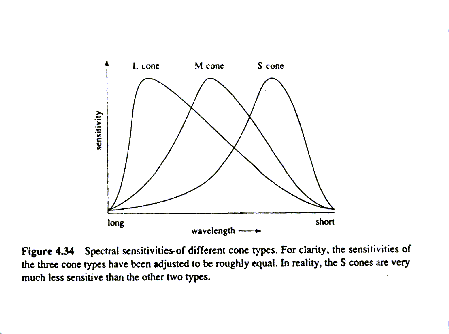
of 3 different cone types to stimulation with monochromatic light of
different colours.
Three different types of cone provide trichromatic vision.
Cones are at highest density in the fovea - rare in periphery.
They have smaller receptive fields than rods - so greater acuity at
fovea, and geater sensitivity (but no colour vision) in periphery.
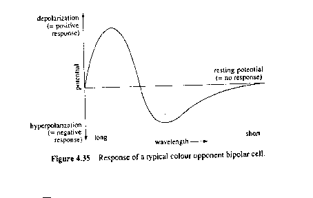
The problem of perceiving colour is solved by colour opponency.
See Figure 4.35 of Book 3 reproduced below.
The term `recognize' means to know again (re-cognize) - implies comparison of incoming information against some internal representation.