|
Here is that fuzzy mess again. If you look closely, you may get the correct impression of a filamentous composition to the material. In fact, we are at the distal surface of the retina of the phalangid eye, just under the lentigen cells which make up the glassy body beneath the lens. The label, prm, indicates the pre-retinal membrane, which is
a rather amorphous structure. You can, however, also see some cellular
features in this image. Remarkably, these are the distal ends of glial
processes, parts of the glial cells which we have already encountered in
May
and June.
|
There's not much to see in this particular
image,
but a somewhat wider view is described in the Opiliones Notes. 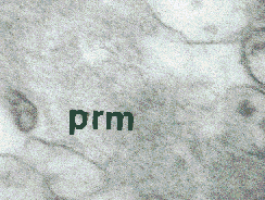 |
|