Cuticle structure

The electron micrograph shown below shows part of the exocuticle in the lens of the eye of Mitopus morio. The lens is a cuticular thickening on the outside of the eye and its structure shows the organisation typical of arthropod cuticles in general.
Although on first glance there appear to be parabolic microfibrils making up the cuticle, this in fact is just an illusion. There are layers in which parallel fibrils run in slightly different directions from those in adjacent layers (lamellae).
This can be seen in the image series below in which you can effectively "dissect out" a set of seven lamellae and see the orientation of their fibrils.

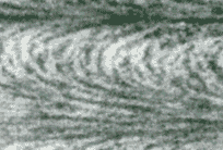
Electron micrograph of the cuticle.

Reset button
Put the layers back and start again.
Slight separation of a few lamellae
1.  Use this button to separate out a few layers in the cuticle
More separation and tilting of a few lamellae
2.  This button will separate out the layers a bit more and tilt them up to give you a clearer view.
Plan view of a few separated lamellae
3.  Finally, the layers are laid out for you in full plan view by this button.
The effect arises because the fibrils in the top and bottom lamellae of our sample are cut in longitudinal section (and show up as elongated profiles), in contrast to those in the centre which are cut transversely (and so are effectively short dots). In between these dots and dashes the intermediate fibrils are cut obliquely.
Thus we get the misleading impression of parabolae.


Back to:-
Lens of the eye Opiliones NotesClick here for Opilio Notes ArachnologiaBack to Arachnologia home page
Back to AriadneAriadne Home Page