March's mystery organelle - amazing!
Once again, the electron micrographs are from of the eye of Mitopus
morio - but all other eukaryotic organisms possess these organelles.
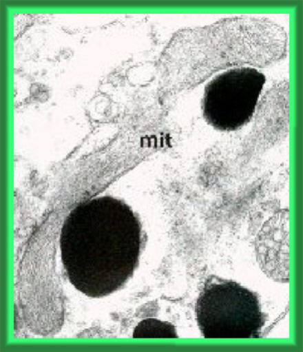
In this enlarged extract from the mystery picture, the object in question
is labelled "mit", which stands for mitochondrion.
This is one kind of organelle found in the cells in the phalangid eye.
Its name comes form the Greek, "mito-" meaning "thread" and "chondrion"
meaning "body", referring to the commonly thread-like shape of these organelles.
I find two aspects of these organelles quite remarkable: the intimate correlation
between their structure and function and their
astounding evolutionary significance.
Perhaps, if you're not a biologist I need to explain (briefly) two of
the terms used so far.
Organelles: Our own bodies, like those of all of the living organisms
that you can see around you in the world (but not some of the micro-organisms
invisible to the naked eye), are made up of cells. We have billions of
these cells in our bodies and one of the important advantages of this is
that different kinds of cells can be specialised for different functions.
Within these cells, we can see smaller structures which, themselves, are
specialised to perform different functions within the cells - these structures
are called organelles ("small organs" - cf. the "organs" which make up
the body).
Eukaryotic: The kind of cells in which we can clearly see
these organelles and, in particular, which have a membrane separating their
nucleus from the surrounding cytoplasm, are termed eukaryotic (from the
Greek meaning "true nucleus").
The electron micrograph is, essentially, just a map of electron density
over a section of the tissue. A beam of electrons is focussed through a
thinly-cut section of the tissue (after it's been preserved, stained with
electron-dense chemiscals and embedded in araldite resin). Where the electrons
pass through unimpeded, the picture shows white; dark (or black) regions
show where the cellular material itself is electron-dense or where it has
picked up the electron-dense stain. Because the section is very thin, it
just cuts throgh the organelles so a single structure can pass in and out
of the section - thus my ambiguity about just precisely how many mitochondria
are in the picture.
Amazing facts of sub-cellular biochemistry
 So
what we have here are the mitochondria (the plural of "mitochondrion").
So
what we have here are the mitochondria (the plural of "mitochondrion").
These organelles are wonderfully organised and structured to be very
efficient in carrying out their role in the cell. Their main role is in
the energy metabolism of the cell. In aerobic respiration, molecules derived
from food (usually) are broken down by oxidation and as part of this process
molecules of a high-energy compound called ATP (adenosine tri-phosphate).
These molecules of ATP can then be used to drive chemical reactions in
the cells which require energy.
Within the mitochondria, the main series of chemical reactions is called
the Krebs cycle (after its initial discoverer, or the TCA cycle - tri-carboxylic
acid cycle - after some of the compounds involved). These reactions depend
upon particular molecules in the membranes of the mitochondria and their
overall structure is geared to this, with infoldings (called cristae) of
the membrane to increase the membrane surface area, as shown in this diagram.
Even more remarkably, within these membranes the molecules are arranged
spatially next to each other for optimal operation!
A good place to find out more about mitochondrial
structure and function in the Cell Biology pages of the University
of Arkansas, where there are also pages on
various aspects of cell structure.
Amazing facts of biological evolution
Even more astounding is the evolutionary origin of these mitochondria.
These days, we think of the millions of living organisms in terms of Five
Kingdoms, of which Plants and Animals are just two. The Fungi are one of
these kingdoms and the other two are different kinds of micro-organisms
(termed Protoctista and Monera by some authors). The prime author describing
these kingdoms is Lynn Margulis, who has written a nice
concise summary for us. This, to some extent has been superseded by
a view of the living world in terms of three domains - Archaea, Bacteria
and Eukarya (= Eukaryota, including plants and animals, etc.). It is quite
easy to appreciate how these classification
systems (5 kingdoms vs. 3 domains) compare.
The really amazing thing here is that the mitochondria were originally
separate organisms, but early in the evolution which led to the eukaryotic
organisms, they moved into the cells of the eukaryotes and became endosymbiotic.
In other words, both the mitochondria and their hosts benefited from their
living within the hosts' cells. The mitochondria even have their own separate
reproduction and genetic systems from those of the organisms in which they
live. As shown in the diagram above, you can see DNA and ribosomes inside
the mitochondria - in other words, they have their own genetic system.
So - we wouldn't exist nor could we survive without these tiny organisms
living symbiotically within our cells - incredible isn't it!!!
You can see more details of the organelles in the phalangid eye
cells in the Opiliones Notes.
Back to Opiliones Notes